




Endometrial Biopsy for Endometriosis and Definitive Tissue Diagnosis
What each biopsy samples, why results can disagree, and when targeted tissue confirmation matters
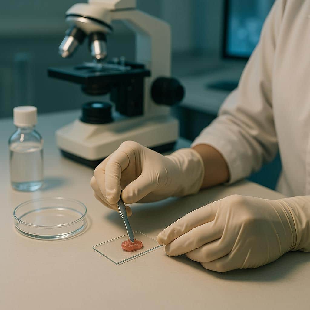
Biopsy confirms endometriosis when it samples the right tissue
An office endometrial biopsy generally cannot confirm endometriosis elsewhere in the pelvis because it samples only the uterine lining. A biopsy taken from a suspected endometriosis lesion can confirm the diagnosis through microscopic tissue examination, called histology. A negative sample does not necessarily exclude disease: the location, depth, and accuracy of sampling matter.
If you’d like a specialist’s view on conflicting biopsy results, imaging, or a recommended treatment, our team can help review your options.
Symptoms, imaging, and surgical inspection can suggest endometriosis, but tissue provides the best confirmation. Laparoscopy and robotic surgery are excellent tools for finding and treating disease, yet appearance alone can both miss endometriosis and mistake other tissue changes for it. A biopsy is most helpful when it answers a specific question, rather than being expected to explain every symptom by itself.
Endometrial and peritoneal biopsies sample different tissues
An office endometrial biopsy samples the uterine lining
An endometrial biopsy takes a small sample from the lining inside the uterus, usually through the cervix. It is used to evaluate abnormalities of that lining. It does not reach suspected endometriosis on the pelvic lining, ovaries, bowel, or other sites outside the uterine cavity.
If you are scheduled for a biopsy for endometriosis, ask exactly which tissue will be sampled and how. An office endometrial biopsy and a surgical biopsy of a pelvic lesion answer different questions. A normal uterine-lining sample does not rule out endometriosis elsewhere.
A peritoneal or lesion biopsy samples suspected disease
During laparoscopy or robotic surgery, the surgeon can remove tissue from a suspicious pelvic lesion and send it to a pathology laboratory. A peritoneal biopsy samples the lining of the abdominal or pelvic cavity. Other lesion biopsies target the particular site where disease is suspected.
Finding the right site requires careful inspection. Endometriosis can be subtle, lie beneath a normal-looking surface, or sit within small peritoneal pockets or windows, sometimes called Allen-Masters pockets. Surgeon experience, camera quality, and implant location all affect whether these areas are found and adequately sampled.
What the tissue examination can establish
The pathologist looks for endometrial-type glands and stroma, the tissue that supports those glands. Bleeding or iron-laden macrophages, cells containing remnants of old blood, may also support the diagnosis.
Tissue examination can confirm endometriosis, challenge a surgeon’s visual impression, or help exclude another condition that resembles it. Depending on the clinical situation, those alternatives may include scar tissue, inflammatory conditions, or some cancers.
Why visual findings and biopsy results can disagree
A visible lesion is not always confirmed on pathology
In a diagnostic-accuracy study of 976 women undergoing laparoscopy for pelvic pain and/or infertility, surgical findings were compared with histopathology. Visual assessment was highly sensitive, meaning it identified most cases confirmed by pathology. It also had a high negative predictive value: when laparoscopy did not suggest endometriosis, disease was usually absent by the study’s reference standard.
Dr. Vasilev cautions that this negative predictive value may be worse in real-world surgery involving non-expert surgeons. Even in that study, specificity and positive predictive value were lower, meaning some areas that looked like endometriosis were not confirmed on pathology.
In a 2021 pilot study of 15 women scheduled for diagnostic laparoscopy for suspected peritoneal endometriosis, surgeons inspected tissue using white light and near-infrared fluorescence. Only about 61% of the suspicious lesions biopsied contained endometriosis on histology. A lack of confirmation does not mean the person’s symptoms were not real; it means visual suspicion and tissue confirmation are different findings.
A negative biopsy may reflect where or how tissue was taken
A negative result needs to be interpreted alongside the operative findings and the tissue sampled. Possible explanations include:
- Sampling error: the biopsy missed a tiny or patchy focus within a larger abnormal area.
- Variation within a lesion: one part contains diagnostic glands and stroma, while another contains mostly inflammation or fibrosis.
- Insufficient depth: a small, superficial sample did not reach deeper disease.
- Look-alikes: scar tissue, inflammation, vascular changes, or other benign findings resembled endometriosis.
- Technical effects: cautery damage or other sampling and tissue-handling factors made interpretation difficult.
Before decisions about long-term hormonal treatment, repeat surgery, or a major bowel or ureter operation, review the pathology report alongside the operative report with your doctor. Ask which locations were sampled, how many samples were taken, whether they reached the suspected disease, and whether pathology confirmed endometriosis or described only nonspecific changes. These details help distinguish an unconfirmed visual impression from a sample that may have missed disease.
Targeted biopsy can clarify bowel or unusual findings
People often seek a diagnosis to explain pain, bleeding, infertility, or gastrointestinal symptoms. In some circumstances, tissue sampling becomes more urgent because clinicians need to exclude cancer or another serious disease. The following examples are uncommon situations, not routine biopsy pathways for pelvic pain.
Bowel disease may be beyond a colonoscopy biopsy
Bowel endometriosis often lies in deeper layers of the bowel wall, beyond the inner lining sampled during routine colonoscopy. A normal surface biopsy therefore does not necessarily rule it out.
A 2023 case report described a 42-year-old woman with a rectal mass causing a large bowel obstruction. She needed a temporary colostomy, and routine endoscopic biopsies showed normal bowel lining, but imaging still raised concern for malignancy. Endoscopic ultrasound identified a deeper lesion, and ultrasound-guided fine needle aspiration obtained tissue that confirmed bowel endometriosis.
For a bowel mass or unexplained narrowing, a normal colonoscopy biopsy may leave the central question unanswered. Endoscopic ultrasound can sometimes identify and guide sampling of disease beneath the surface.
Possible bowel obstruction needs emergency evaluation. If you have severe abdominal pain or swelling with vomiting or an inability to pass stool or gas, call 911 or go to the nearest emergency department.
Conflicting Biopsy Results? Get a Specialist’s Perspective
Our specialists are here to help you understand your condition and explore your treatment options.
Schedule a ConsultationAn unusual peritoneal finding may offer a biopsy target
A 2019 case report described a 24-year-old woman with recurrent, massive hemorrhagic ascites, meaning blood-stained fluid accumulating in the abdomen. She also had anemia, menstrual pain, and an elevated CA-125 level, findings that can raise concern for malignancy.
Ultrasound eventually identified thickened peritoneal tissue. An ultrasound-guided core needle biopsy of that area confirmed endometriosis. Medical treatment subsequently improved her symptoms and laboratory results, although fluid accumulation had not completely resolved.
This presentation is very rare. Its practical relevance is that a distinct abnormal area on imaging may provide a useful target for tissue sampling when more general testing has not explained the problem.
Nonsurgical sampling is an option only in select cases
Endoscopic ultrasound-guided needle sampling may reach deep rectal or rectosigmoid lesions from inside the bowel. Image-guided core needle biopsy may be possible when an abnormal area is visible and safely accessible through the skin.
These approaches can be valuable when malignancy is a concern, when a deep lesion remains unexplained after normal surface biopsies, or when surgery is high-risk or deferred and tissue confirmation would change treatment. They do not replace laparoscopy for most people with pelvic symptoms and nonspecific imaging.
Needle sampling has limitations and risks. A needle can miss disease by only a few millimeters, and needles introduced through the skin can cause complications. The treating team needs to weigh the safety of reaching the target against how much the result would clarify care.
Better visualization can help find lesions but cannot replace histology
Near-infrared fluorescence remains an investigational aid
Near-infrared fluorescence uses a special camera after intravenous indocyanine green dye to highlight tissue patterns that may differ from surrounding areas. In the 2021 pilot study, it was feasible, and no complications related to the imaging were observed in that small sample.
Fluorescence identified six additional suspicious areas beyond those seen with white light, but only two contained endometriosis. Its added value over standard white-light laparoscopy was limited, and it also produced false alarms.
These tools remain investigational aids to lesion detection. They do not replace histology, and they should not be assumed to prevent missed disease, reduce recurrence, or reduce repeat surgery. Proof of those long-term benefits remains limited.
Camera optics and surgical inspection still matter
Better optics are available now. Conventional laparoscopy usually provides a two-dimensional view, while robotic surgery provides a three-dimensional, more magnified view.
Dr. Vasilev’s view is that robotic optics can make subtle tissue changes clearly visible that may not be apparent with two-dimensional laparoscopy. This can remain true even when a laparoscopist uses “contact laparoscopy,” bringing the camera extremely close for inspection. Better visualization helps the surgeon identify where to sample, but the tissue still needs microscopic examination for confirmation.
Adenomyosis calls for a different tissue strategy
A lining sample may miss disease in the uterine muscle
For adenomyosis, the tissue of concern is within the uterine muscle, beyond the reach of a routine office endometrial biopsy. It can infiltrate that muscle in a diffuse, spiderweb-like pattern rather than form one easily sampled spot. A normal lining biopsy therefore cannot reliably exclude adenomyosis.
Symptoms and imaging, especially transvaginal ultrasound or MRI, often guide diagnosis in routine care. Historically, definitive tissue confirmation came from examining the uterus after hysterectomy, but hysterectomy is neither appropriate nor desired for many patients.
Outpatient hysteroscopy allows inspection inside the uterine cavity and may permit targeted sampling. However, it may or may not obtain tissue deep enough to definitively prove adenomyosis.
Laboratory biopsy research does not change today’s diagnosis
A 2024 laboratory study examined surgical biopsy samples from patients with adenomyosis, ovarian and other genital endometriosis lesions, pelvic peritoneal endometriosis, and controls. It measured oxygen consumption and metabolic gene activity. Adenomyosis samples showed higher cellular respiration, while ovarian and peritoneal lesion samples showed reduced oxygen use and patterns consistent with greater reliance on breaking down glucose for energy.
This testing is not part of routine diagnosis. It reinforces that endometriosis and adenomyosis are related but not identical, and that disease biology varies by lesion type and location. Future non-hormonal treatments aimed at cellular energy use may need to account for those differences rather than use one strategy for every lesion.
Questions to take to your own doctor
If a biopsy is planned, or your surgical findings and pathology results seem to conflict, these questions can help clarify what the test can establish:
- Were biopsies taken of the suspected lesions? How many, and from which locations?
- Did pathology confirm endometriosis, or were the results nonspecific?
- If pathology was negative, could sampling depth or location explain it, particularly with superficial versus deep disease?
- Do my symptoms suggest deep bowel or ureter disease, and do we need targeted imaging or a different sampling approach?
- If there is a bowel mass or stricture, could endoscopic ultrasound-guided sampling help resolve the uncertainty?
For questions about a procedure you have already had, review the results and follow-up plan with the surgeon or care team who treated you. If the pathology, imaging, and proposed treatment still do not fit together, a specialist second opinion can help assess what has been established and what remains unanswered.
References
Toniyan, Malkov, Biryukov et al. The Cellular Respiration of Endometrial Biopsies from Patients with Various Forms of Endometriosis. International Journal of Molecular Sciences. 2024.. DOI: 10.3390/ijms25073680
de Almeida, de Oliveira, do Amaral. Accuracy of laparoscopy for assessing patients with endometriosis. São Paulo Medical Journal. 2008.. DOI: 10.1016/j.fertnstert.2025.12.021
Carvalho, Cardoso, Pires et al. Diagnosis of Bowel Endometriosis Using Endoscopic Ultrasound-guided Fine Needle Aspiration. The Korean Journal of Gastroenterology. 2023.. DOI: 10.4166/kjg.2022.104
Al-Taher, van den Bos, Terink et al. Near-Infrared Fluorescence Imaging for the Intraoperative Detection of Endometriosis: A Pilot Study. Life. 2021.. DOI: 10.3390/life12010015
Wang, Li, Tong et al. Endometriosis presenting with recurrent massive hemorrhagic ascites and diagnosed by core needle biopsy. Medicine. 2019.. DOI: 10.1097/MD.0000000000015477
Quick Answers
Why do I have painful urination and pelvic cramping between periods?
Painful urination and pelvic cramping between periods can happen when the bladder, uterus, pelvic floor, or nerves are irritated. Endometriosis can contribute, but bladder pain syndrome, pelvic floor overactivity, or adenomyosis can also cause these symptoms.
When is hysterectomy recommended for adenomyosis?
Hysterectomy is typically considered for adenomyosis when severe symptoms come from the uterus and you’re not planning future pregnancy. It is usually weighed most strongly when conservative treatments haven’t provided acceptable relief, aren’t tolerated, or don’t fit your goals.
Which adenomyosis symptoms most affect daily life?
Heavy or prolonged periods and severe period pain usually cause the biggest disruption to daily life with adenomyosis. Bleeding can affect work, school, travel, and sleep, while fatigue, pelvic pressure, and pain with sex for some people can add to the burden.
Why does ovarian cyst pain keep coming back?
Ovarian cyst pain can return because new cysts form with ovulation, a cyst persists or comes back, or nearby endometriosis or scar-like bands cause pain. An endometrioma, a cyst caused by endometriosis, may be associated with deeper pelvic disease.