



Deep Infiltrating Endometriosis: Current and Future Treatments
From molecular differences and symptom recognition to diagnosis, medical therapy, advanced surgical techniques, and what’s ahead.
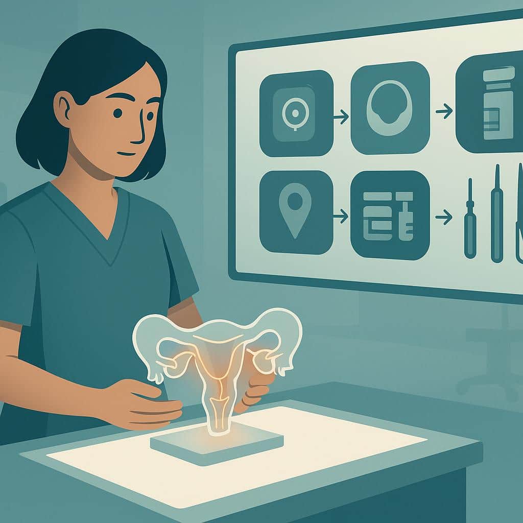
How to Treat Deep Infiltrating Endometriosis
Endometriosis, already challenging when endometrial-like cells grow outside the uterus and on the surfaces of other organs, has a more troubling variant known as deep infiltrating endometriosis (DIE). This severe form is defined by abnormal cells burrowing or invading more than 5 mm below the tissue surface into affected organs such as the bowel and bladder. This guide outlines current approaches to managing DIE and how future advances may improve treatment. At this time, a long-standing cure remains out of reach, even for superficial endometriosis.
Understanding Deep Infiltrating Endometriosis
This condition is relatively uncommon, affecting about 1% of women of reproductive age and about 20% of those with endometriosis. Molecular data suggest it may be a premalignant disease, along with the endometrioma type; research is ongoing, and the malignancy potential remains low. Shared genetic mutations with aggressive diseases like cancer may help explain why endometrioma and DIE types are more likely to cause local anatomic distortion and harm, including pain and subfertility, and why they may have increased metastatic potential even without any associated cancer.
Anatomically, three major types of endometriosis are recognized based on location within the pelvic and abdominal cavities. Superficial endometriosis (also called peritoneal endometriosis or PEM) involves lesions appearing on the surface of organs outside the endometrium and generally, though not always, causes the least tissue damage and distortion. Ovarian endometriomas (OE) are dark cysts caused by old collected blood, commonly called chocolate cysts, that form in or on the ovaries due to endometriosis. Deep infiltrating endometriosis is the most severe form, in which endometrial-like tissue penetrates deeply into pelvic organs and causes more extensive damage.
Current staging systems rely largely on anatomic location and clinical degree of disease. This approach is considered “old school,” and a new era is emerging in which molecular insights are expected to improve diagnostics and treatment because abnormal molecular pathways can be targeted with precision therapies.
Molecular Comparison of Endometriosis Types
Research in this area extends well beyond hormonal factors such as receptor activity, regulatory changes, and relative progesterone resistance. Although it is too early to classify different types of endometriosis strictly by gene mutation pathways, what is known may already help with recurrence and cancer risk mitigation. In summary, multiple genes can be mutated in endometriosis, which may or may not lead to a low risk of cancer but can influence how aggressive DIE and OE are in an individual. These gene mutations may be suppressed or aggravated by epigenetic influences such as diet, lifestyle, toxin exposure, concurrent disease states, and mind–body factors. Ongoing research is uncovering diagnostic possibilities and molecularly guided therapies based on these findings.
Identifying Symptoms of Deep Infiltrating Endometriosis
Symptoms are similar to those of general endometriosis but are usually more severe. They can include severe pelvic pain, painful urination (dysuria) or blood in the urine (hematuria), painful menstruation (dysmenorrhea), genital pain before, during, or after sex (dyspareunia), digestive discomfort and rectal bleeding, and distant symptoms such as pain with breathing when the diaphragm is involved.
Causes and Risk Factors of Deep Infiltrating Endometriosis
Despite recent advances, the exact cause of endometriosis, including DIE, remains unknown. Family history of endometriosis and/or cancer is important to consider. Because the condition is likely multifactorial, a single cause is unlikely to be identified, and molecular research is pushing understanding to a deeper level.
Diagnosing Deep Infiltrating Endometriosis
As an advanced form of endometriosis, DIE can be challenging to diagnose. Multiple diagnostic modalities are typically employed, including medical history, physical examination, histological evaluation after surgery or biopsy (such as evaluation of a C-section scar endometriosis), minimally invasive surgery, ultrasound, and MRI. A 3-Tesla (“3T”) MRI is probably the most accurate imaging modality, yet it still has significant limitations. It can aid in surgical planning but should not be used to determine definitively whether DIE is present or absent, as it can miss up to 20% of DIE.
In many—if not most—cases, the diagnosis is only apparent and confirmed during surgery. Because it is impossible to precisely predict the full extent of disease beforehand, choosing the most skilled and experienced surgeon is prudent, particularly when symptoms are severe. A poorly executed surgery complicates subsequent procedures and increases the risk of major complications.
Treatment Options for Deep Infiltrating Endometriosis
Medical Treatments
Medical therapy for DIE is extremely limited and essentially non-existent in terms of disease eradication. Deep invasive disease triggers scarring or fibrosis as the body attempts to wall off and heal the area, and no known medication can eliminate fibrosis. Pain relievers and hormonal therapies used for other forms of endometriosis may provide symptomatic relief and possibly some suppressive effect.
Integrative strategies can help with symptom control in DIE much as they do with other types of endometriosis. Mind–body–based biofeedback, nutrition, botanicals, essential oils, acupuncture and acupressure, and electrical stimulation (TENS) are examples, and it is best to craft such an approach with a qualified practitioner.
Pelvic floor physical therapy (PFPT) plays a central role in treatment planning. Depending on lesion location, caution is advised with internal therapies because disruption of deep lesions can potentially make surgery less effective. A coordinated team approach is ideal to determine the best strategy.
Take Control of Deep Endometriosis Today
Our specialists are here to help you understand your condition and explore your treatment options.
Schedule Your ConsultationSurgical Treatments
Surgical excision of DIE lesions and associated fibrosis is the best path forward in most cases where DIE is suspected or has been diagnosed previously. The usual emphasis on excision over ablation is even more crucial here because ablation or fulguration is not useful for lesions of uncertain depth and carries an elevated risk of damaging structures such as the rectum, bladder, and ureters.
A master surgeon is best suited for DIE. In the author’s opinion, these procedures should be performed robotically because of superior optics and wristed robotic instruments. The surgeon should be prepared to address bowel, bladder, and ureteral involvement, including reimplantation when necessary, or should work with a well-integrated team ready to participate as planned. Although it is difficult to predict all requirements preoperatively, careful imaging-based mapping and close attention to symptoms improve preparation for comprehensive resection or excision.
DIE does not automatically necessitate a hysterectomy. If childbearing is complete, the risks and benefits of hysterectomy may need to be discussed to ensure all diseased tissue is removed. As menopause approaches, and with more extensive disease or a higher malignancy risk due to family history or genetic testing, it becomes more prudent to weigh the risks and benefits of ovarian conservation. Decisions should be individualized and thoroughly discussed.
Considerations in Surgical Management
Indocyanine Green (ICG) Fluorescence
Indocyanine green (ICG) fluorescence imaging helps surgeons visualize the ureters and safely remove as much infiltrating endometrial tissue as possible without damaging the urinary tract. It is also valuable for assessing blood supply and viability in bowel segments that have been operated upon, reducing the risk of complications.
Stenting During Partial Cystectomy or Ureteral Reimplantation
During bladder surgery for urinary endometriosis, the placement of stents—tiny plastic catheters—can protect the ureters and enhance healing after reimplantation.
Pathology Evaluation
Beyond standard pathology, newer assessments may be considered in DIE and OE, especially when disease appears clinically aggressive. The pathologist should ensure there is no clear cell cancer component. Even without cancer, tissue can be evaluated for mitotic index to determine how many cells are dividing, a finding more common in aggressive disease and one that may prompt consideration of well-tolerated hormone suppression such as micronized progesterone to reduce recurrence risk. Immunohistochemical (IHC) stains can assess molecular abnormalities driven by gene mutations, such as ARID1A; while not readily available, they may be useful when cancer risk is elevated. In the future, these aberrant molecular pathways are expected to be targeted with precision therapies.
Endometriosis and Fertility
Endometriosis, including DIE, can affect fertility. Surgeons should prioritize atraumatic techniques to protect delicate reproductive structures and improve pregnancy prospects, a goal optimized with the robotics platform.
Key Molecular Facts at a Glance
- Endometriosis overall (PEM, OE, and DIE types) shows genetic and molecular heterogeneity, with multiple genes affected. Some mutations mirror those found in cancer even when no cancer is present or likely to develop. This suggests endometriosis may represent different disease entities between individuals, with varying aggressiveness. Within a single individual, research supports clonal molecular signature similarities between PEM, OE, and DIE, indicating that one type can progress to another.
- Mutations of interest include ARID1A, PIK3CA, KRAS, and PPP2R1A, among others.
- The endometrioma (OE) type carries the highest risk of malignant degeneration, and ARID1A is considered a key driver mutation for clear cell cancer.
- DIE has a broader range of mutations and a lower risk of malignant degeneration than OE, but these mutations may contribute to more aggressive behavior.
- OE may have a higher malignant degeneration risk than DIE because the ovarian microenvironment may be more permissive to malignant change; this does not negate the aggressive, invasive, and potentially metastatic nature of DIE in the absence of cancer.
- Gene mutation expression varies under epigenetic influences, including diet, lifestyle, toxin exposure, concurrent disease states, and mind–body factors.
Quick Reference: Prevalence and Imaging Performance
- Prevalence among reproductive-age women: about 1%
- Proportion among all endometriosis cases: roughly 20%
- 3-Tesla (3T) MRI: currently the most accurate imaging option, though it can still miss up to 20% of deep infiltrating endometriosis (DIE), so it shouldn’t be used as definitive proof for or against disease.
Conclusion
Deep infiltrating endometriosis adds complexity to the management of endometriosis. Surgery is the optimal therapy, followed by supportive care and strategies to mitigate recurrence where possible. Malignant degeneration is uncommon but possible, and with a family history of cancer, genetic testing should be considered. The more complex the situation, the greater the need for an endometriosis expert master surgeon. If cancer risk is elevated for any reason noted here, consultation with a gynecologic oncologist should be considered.
References
The Diagnosis and Treatment of Deep Infiltrating Endometriosis DOI: 10.1016/j.gore.2025.101985
Quick Answers
How is multi-organ endometriosis treated without organ removal?
In many multi-organ cases, the goal is conservative surgery: removing endometriosis while preserving the organs themselves. That typically means meticulous excision of disease from surfaces and deeper planes around the bowel, bladder, ureters, ovaries, and pelvic sidewalls—freeing organs from scar tissue and restoring normal anatomy without taking the organ out. Because endometriosis can hide in distorted or “frozen” anatomy, the safest way to preserve organs is often a highly precise approach that can dissect around vital structures.
In our practice, we use robotic excision to improve visualization and fine-control dissection, which is especially helpful when disease involves multiple compartments or has been operated on before. When endometriosis is close to structures like the ureters, bowel, diaphragm, or certain nerves, treatment planning may include coordinated work with other surgical specialists so the disease can be fully addressed in one operation while still prioritizing organ-sparing techniques.
Organ removal is usually considered only when an organ is severely damaged, there are multiple large endometriomas that can’t be safely managed with tissue-sparing techniques, fertility-safety concerns arise (like a badly damaged tube), or there’s concern for tumor or malignant change. If you’re trying to avoid organ removal, we can help map likely disease sites, clarify your priorities (pain relief, function, fertility), and outline what organ-preserving excision could realistically look like in your specific case—then build a surgical plan around that.
How much time off work do I need after endometriosis surgery?
Most people need about 2–3 weeks off work after minimally invasive endometriosis excision, especially if your job is mainly desk-based and you can ease back in. With robotic excision, patients often go home the same day or next day, start walking comfortably within about a week, and many feel ready for a gradual return to typical daily routines in that 2–3 week window.
The exact time off depends less on the incision size and more on what we need to treat during surgery—for example, ovarian endometriomas, bowel/bladder/ureter involvement, extensive scar tissue (“frozen pelvis”), or additional procedures like appendix removal or adenomyosis-related surgery. More complex, multi-organ cases can mean more fatigue, more activity restrictions, and a higher chance of needing an overnight stay, which can extend the time you’ll want to plan away from work.
In most straightforward recoveries, many patients are back to full activity by about a month. If you tell our team what you do for work (lifting, long shifts, travel, on-your-feet all day vs remote/desk), we can help you plan a realistic time-off request and a safer return-to-work ramp based on the surgical plan we’re building for you.
When is hysterectomy recommended for adenomyosis?
A hysterectomy is typically considered for adenomyosis when symptoms are severe and clearly uterus-driven—most often heavy bleeding (sometimes with anemia), intense cramping, pelvic pressure, and daily quality-of-life disruption—and you’re not planning future pregnancy. It’s the most definitive option because adenomyosis lives within the uterine muscle, so removing the uterus removes the source of the problem.
In practice, we usually weigh hysterectomy most strongly when conservative options haven’t brought acceptable relief, aren’t tolerated, or don’t fit your goals. The decision also depends on the pattern and extent of disease (diffuse adenomyosis versus a more focal adenomyoma that may be removable while preserving the uterus) and whether endometriosis may also be present. If endometriosis is part of the picture, it’s important to know that hysterectomy alone doesn’t treat disease outside the uterus—durable symptom relief depends on addressing all pain generators.
If you’re wondering whether you’re at the point where hysterectomy makes sense, our team can help clarify what’s most likely driving your symptoms, review imaging, and walk you through uterus-preserving versus definitive surgical paths so you can choose the option that best matches your relief and fertility priorities.
Why does ovarian cyst pain keep coming back?
Ovarian cyst pain can feel “recurrent” for a few different reasons. Some cysts are functional (they form with ovulation and then resolve), so the pain returns in a similar spot month after month even though it’s not the exact same cyst. In other cases, the cyst itself can come back or persist—especially if it’s an endometrioma (an ovarian cyst caused by endometriosis), which can behave differently than a simple cyst and may be associated with deeper pelvic disease.
Another common reason is that the cyst isn’t the whole story: endometriosis on the pelvic sidewall, uterosacral ligaments, bowel, bladder, or around the ovary can irritate the same nerves and tissues and make it feel like “my cyst is back” when the driver is actually inflammatory disease nearby. Adhesions (scar-like bands) can also tether the ovary and cause recurring pulling or sharp pain, even when imaging doesn’t show a large cyst. If your pain cycles, escalates, returns quickly after a “normal” ultrasound, or keeps recurring despite prior treatment, our team can help you map the pattern, interpret imaging with an endometriosis lens, and decide whether targeted evaluation and—when appropriate—excision surgery is the next best step.
What is deep infiltrating endometriosis (DIE) and how is it treated?
Deep infiltrating endometriosis (DIE) is endometriosis that grows deeper into tissue—often described as more than ~5 mm below the surface—and it commonly involves structures like the uterosacral ligaments, rectovaginal space, bowel, bladder, or ureters. Because it can inflame, scar, tether, or even narrow/obstruct nearby organs, DIE may show up as “non-gynecologic” symptoms such as bowel or urinary pain, painful sex, nerve-type pelvic pain, or symptoms that don’t match a routine pelvic exam.
Treatment depends on where the disease is and what it’s affecting, but DIE is the subtype most likely to require specialized surgical planning. When surgery is appropriate, meticulous excision (removing disease at its roots rather than burning the surface) is the gold-standard approach for durable symptom relief and for addressing organ involvement; in complex cases this can include careful work around the bowel, bladder, and ureters. Our team focuses on advanced, precision excision (often using robotic technology) with a tailored plan that prioritizes safety, completeness, and your goals—whether that’s pain relief, fertility, or protecting organ function.
Because DIE can be missed on basic exams and even “normal” imaging, evaluation often requires a higher index of suspicion and the right strategy for mapping disease before any procedure. If DIE sounds like it could explain your symptoms, explore our detailed resources on deep disease and excision, or reach out to schedule a consultation so we can review your history, imaging, and next steps together.