



Do You Need Two Ultrasounds Before Bowel Endometriosis Surgery?
Mapping rectal endometriosis with TVS and ERUS to tailor surgery and expectations
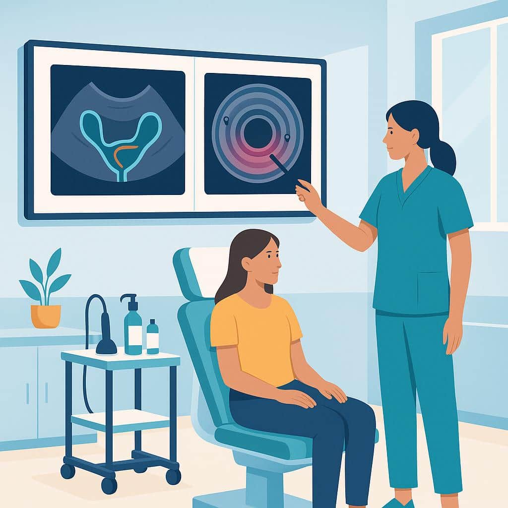
When rectal endometriosis is suspected, “mapping” changes everything
If you’re living with bowel symptoms—pain with bowel movements, constipation/diarrhea flares around your period, deep pelvic pain, painful sex, bloating, or that frightening feeling of “something is stuck”—it can be validating (and overwhelming) to hear the words rectal endometriosis or bowel deep endometriosis.
One of the biggest practical challenges is that bowel endometriosis isn’t just “present or not.” What matters for your treatment plan is where it is, how big it is, and how deep it goes into the bowel wall—because that can change the kind of surgery recommended, the surgeon you need on the team, and your risk of bowel complications.
Recent evidence supports a very patient-relevant idea: using two different ultrasound approaches before surgery can give a fuller picture than using only one—especially when the goal is planning the safest, least aggressive surgery that still treats your symptoms.
The two ultrasound tests you may hear about
Transvaginal ultrasound (TVS)
This is the pelvic ultrasound done with a vaginal probe (often similar to scans used in fertility workups). Many endometriosis-experienced sonographers can “map” deep endometriosis with TVS—looking at ovaries, uterosacral ligaments, pouch of Douglas, and signs that bowel may be involved.
Real-world plus: It’s widely available and can assess pelvic organs beyond the bowel.
Common limitation: TVS may not reliably show all rectal nodules or clearly define which bowel wall layers are involved.
Endorectal ultrasound (ERUS)
ERUS uses a probe in the rectum to visualize the rectal wall in layers. If you’ve been told, “We need to know whether this goes into the muscular layer or deeper,” ERUS is one tool that aims to answer that.
Real-world plus: It can give detailed information about depth of invasion in the rectal wall.
Common limitation: It may sometimes overestimate how deep disease goes (which matters because “deeper” can push surgeons toward more extensive procedures).
What “two ultrasounds” can add for you
In a small prospective cohort where patients had both TVS and ERUS before surgery for rectal endometriosis, TVS identified rectal nodules in about 60.9% of patients, while ERUS characterized nodules in all included patients. In other words: if TVS doesn’t clearly show a rectal nodule but your symptoms and exam strongly suggest bowel involvement, ERUS (in the right hands) may provide additional detail.
The more important patient-facing point isn’t “which test is best” in the abstract—it’s this:
Better pre-op mapping can help your team plan a surgery that fits your anatomy, including whether bowel surgery is likely and what type. It can also help you prepare emotionally and practically (time off work, recovery expectations, the right surgical specialists in the room).
Why depth and “layers” matter (and why ERUS can be helpful)
Your rectum has layers. Endometriosis might affect:
- the outer surface,
- the muscular layer (common in deep disease),
- and rarely the submucosa/mucosa (deeper layers closer to the inside).
In this cohort, ERUS was reported to have 100% sensitivity for detecting mucosa/submucosa involvement (meaning it caught all cases that truly had it), but 73.9% specificity (meaning it sometimes suggested mucosal/submucosal involvement when surgery/pathology didn’t confirm it). For you, that translates to:
- ERUS can be useful for not “missing” deep involvement.
- But if ERUS suggests very deep invasion, it’s worth asking how your team confirms that finding and how strongly it will influence the surgical plan. If the team includes surgeons who are used to performing resections and stapled anastomosis (with or without a colostomy being required, which is usually temporary but lifestyle altering), then you may not get consideration of a lesser resection like a disc or even shaving attempt first.
How imaging can influence the type of bowel surgery offered
If bowel endometriosis is confirmed and you’re considering surgery, there are several surgical approaches. The goal is symptom relief while minimizing risk.
In this cohort, surgeons most often used:
- Rectal shaving: 52.2% (12/23)
- Disc excision: 34.8% (8/23)
- Segmental resection: 13.0% (3/23)
Here’s what those terms generally mean in patient language:
Shaving: Removing endometriosis from the surface of the bowel (and sometimes into superficial muscle) without cutting out a full thickness segment. Often considered “less aggressive.”
Disc excision: Removing a “disc” of bowel wall where the nodule is (full-thickness at that spot) and closing the bowel.
Segmental resection: Removing a segment of bowel and rejoining the ends. This is usually considered when disease is extensive, involves multiple areas, causes narrowing, or deeply infiltrates in a way that shaving/disc excision may not be safe or effective.
Get Accurate Bowel Endometriosis Imaging
Our specialists are here to help you understand your condition and explore your treatment options.
Schedule Your ScanA key practical detail: narrowing (stenosis) showed up with segmental resections
In this cohort, luminal stenosis (narrowing) was seen in 2 out of 3 people who had segmental resection (66.7%)—and those patients also had larger nodules on ERUS measurements.
You shouldn’t take that as “segmental resection causes stenosis” (the numbers are tiny and people getting resections are often the most severe cases). But you can take this to your appointment as a real-life planning point:
If imaging suggests a larger lesion or significant narrowing, your surgeon may recommend segmental resection—and you deserve a clear explanation of why, what alternatives exist, and what the functional risks are (bowel habit changes, urgency, constipation, leaks, etc.). In some cases, it may be prudent to have a gastroenterologist perform a colonoscopy to confirm what they see and to "ink" areas that look like they are involved very deeply or even into the lumen (the inside) of the bowel.
What you might feel better after surgery—and what might not change quickly
At 3 months after surgery in this cohort, multiple endometriosis quality-of-life areas improved significantly, including:
- Pain
- Emotional well-being
- Work-related functioning
- Sexual relationship concerns
That matches many patients’ lived experiences: pain relief can be meaningful, and the mental load can lift when symptoms are treated.
But digestive quality-of-life (measured by a GI quality-of-life tool) improved only numerically and was not statistically significant at 3 months. A very practical takeaway is:
Your bowel function may take longer to settle than your pelvic pain.
It’s also possible that some bowel symptoms come from overlapping issues (IBS, pelvic floor dysfunction, dietary triggers, nerve sensitization) even when endometriosis is treated well.
If bowel symptoms are your main complaint, talk with your team about a recovery timeline that includes pelvic floor rehab, diet support, and realistic expectations for the first 3–6 months.
Safety: what complications looked like here
In this cohort, there were:
- No Grade III or IV complications reported
- One Grade II complication (rectal bleeding)
This is reassuring, but it’s still crucial to personalize risk. Complication rates depend heavily on:
- surgeon and center experience,
- extent of disease,
- whether resection is needed,
- whether you also have bladder/ureter involvement,
- and your baseline bowel function.
Who might benefit most from adding ERUS to TVS?
You might consider asking about ERUS (in addition to TVS) if:
- your symptoms strongly suggest bowel involvement but TVS is unclear,
- your TVS shows a rectal nodule and your surgeon needs more detail on depth,
- there’s concern for bowel narrowing/stenosis,
- or you’re trying to avoid an overly aggressive bowel surgery and want the best possible mapping.
ERUS is not available everywhere and is operator-dependent. In some centers, MRI may be used instead of or alongside ERUS/TVS. What matters is not the “perfect test,” but getting high-quality imaging interpreted by people who routinely map deep endometriosis.
Questions to ask your doctor (bring these to your pre-op visit)
- “Based on my symptoms and exam, do you suspect rectal endometriosis—and what imaging best maps it in your practice?”
- “Would TVS plus ERUS add information in my case, or would MRI be more useful?”
- “If ERUS suggests deep layers are involved, how do you confirm that—and how will it change the surgical plan?”
- “Given my imaging, what’s the most likely procedure: shaving, disc excision, or segmental resection—and what would make you switch plans during surgery?”
- “Will a colorectal surgeon or a gynecologic oncologist be present? If not, under what circumstances would you involve one?”
- “What bowel function changes are most common after the type of surgery you’re recommending, and what support do you offer (pelvic floor PT, dietitian, follow-up plan)?”
Reality check: what this doesn’t settle (and why your plan must be individualized)
Even though dual ultrasound mapping is promising, there are still major unknowns:
- This evidence comes from a small cohort, and imaging performance numbers (like sensitivity/specificity for deep layers) are based on very few cases.
- Follow-up was short (3 months), so it doesn’t answer long-term questions like recurrence, long-term bowel function, or fertility outcomes.
- Imaging quality varies hugely by operator and equipment; a “normal” scan does not always equal “no disease.”
Your best next step is to use imaging as a tool for shared decision-making: balancing symptom severity, your fertility goals, your tolerance for risk, and the experience level of the surgical team.
References
Yan H, Zhang G, Zhong G, et al. Dual ultrasound combination improves the accuracy of preoperative assessment in rectal endometriosis: a prospective cohort study. Annals of Medicine and Surgery. 2025. DOI: 10.1097/MS9.0000000000004293
Quick Answers
How is multi-organ endometriosis treated without organ removal?
In many multi-organ cases, the goal is conservative surgery: removing endometriosis while preserving the organs themselves. That typically means meticulous excision of disease from surfaces and deeper planes around the bowel, bladder, ureters, ovaries, and pelvic sidewalls—freeing organs from scar tissue and restoring normal anatomy without taking the organ out. Because endometriosis can hide in distorted or “frozen” anatomy, the safest way to preserve organs is often a highly precise approach that can dissect around vital structures.
In our practice, we use robotic excision to improve visualization and fine-control dissection, which is especially helpful when disease involves multiple compartments or has been operated on before. When endometriosis is close to structures like the ureters, bowel, diaphragm, or certain nerves, treatment planning may include coordinated work with other surgical specialists so the disease can be fully addressed in one operation while still prioritizing organ-sparing techniques.
Organ removal is usually considered only when an organ is severely damaged, there are multiple large endometriomas that can’t be safely managed with tissue-sparing techniques, fertility-safety concerns arise (like a badly damaged tube), or there’s concern for tumor or malignant change. If you’re trying to avoid organ removal, we can help map likely disease sites, clarify your priorities (pain relief, function, fertility), and outline what organ-preserving excision could realistically look like in your specific case—then build a surgical plan around that.
Why do I look pregnant from bloating with constant pelvic pressure?
Feeling so bloated you “look pregnant” along with constant pelvic pressure usually points to more than simple gas—often it’s a pelvic condition creating inflammation, swelling, or a sense of bulk. Endometriosis can irritate the bowel and pelvic lining, trigger scarring that tethers organs, and create the classic “endo belly” sensation that comes and goes (sometimes not perfectly cyclical). Pelvic pressure can also happen when endometriosis involves deeper tissues or nearby organs like the bladder, ureters, or rectum.
Just as important: these symptoms can be driven by endometriosis neighbors or coexisting conditions, especially adenomyosis and fibroids, which can make the uterus feel heavy, full, or “bulky” and add pressure on the bladder and bowel. Ovarian cysts and other benign pelvic findings can contribute, and IBS-like bowel sensitivity can overlap so closely that symptoms alone don’t reliably sort out what’s causing what. Our team focuses on mapping the full picture—uterus, ovaries, bowel, bladder, and pelvic support structures—so treatment targets the true driver(s), not just the most obvious diagnosis.
If this pressure/bloating is persistent, worsening, or changing your ability to eat, move your bowels, or urinate comfortably, it’s a strong reason to pursue a deeper evaluation rather than being told it’s “normal.” You can explore our educational content on bowel symptoms, bladder symptoms, and overlapping conditions, and reach out to schedule a consultation so we can review your history, imaging, and symptom pattern and outline a plan aimed at lasting relief.
What is deep infiltrating endometriosis (DIE) and how is it treated?
Deep infiltrating endometriosis (DIE) is endometriosis that grows deeper into tissue—often described as more than ~5 mm below the surface—and it commonly involves structures like the uterosacral ligaments, rectovaginal space, bowel, bladder, or ureters. Because it can inflame, scar, tether, or even narrow/obstruct nearby organs, DIE may show up as “non-gynecologic” symptoms such as bowel or urinary pain, painful sex, nerve-type pelvic pain, or symptoms that don’t match a routine pelvic exam.
Treatment depends on where the disease is and what it’s affecting, but DIE is the subtype most likely to require specialized surgical planning. When surgery is appropriate, meticulous excision (removing disease at its roots rather than burning the surface) is the gold-standard approach for durable symptom relief and for addressing organ involvement; in complex cases this can include careful work around the bowel, bladder, and ureters. Our team focuses on advanced, precision excision (often using robotic technology) with a tailored plan that prioritizes safety, completeness, and your goals—whether that’s pain relief, fertility, or protecting organ function.
Because DIE can be missed on basic exams and even “normal” imaging, evaluation often requires a higher index of suspicion and the right strategy for mapping disease before any procedure. If DIE sounds like it could explain your symptoms, explore our detailed resources on deep disease and excision, or reach out to schedule a consultation so we can review your history, imaging, and next steps together.
Why are bowel movements painful and bloating worse during my period?
Painful bowel movements and bloating that flare during your cycle are common in endometriosis—especially when disease is deep in the pelvis near the rectum/rectosigmoid, or when inflammation and scarring tether the bowel to nearby structures. Even without endometriosis growing “inside” the bowel, pelvic lesions can irritate surrounding tissue and nerves, creating cramping, pressure, and the deep, sharp pain some people feel with passing stool. Hormonal cycling can amplify inflammation and swelling, which is why symptoms often peak around bleeding and then ease. Many patients also notice alternating constipation/diarrhea or an “endo belly” pattern that tracks with their period.
These symptoms are often confusing because standard GI workups (including colonoscopy) can be normal—bowel endometriosis frequently affects the outside of the bowel wall or deeper layers rather than the inner lining that a colonoscopy evaluates. What matters most is mapping where symptoms point anatomically and whether there are red flags like cyclical rectal bleeding, escalating severity, or signs of narrowing/obstruction. Our team focuses on a whole-pelvis evaluation and, when appropriate, targeted imaging and surgical planning to confirm what’s driving your bowel pain and bloating and treat it effectively. If you’d like, you can reach out to schedule a consultation so we can review your symptom pattern and discuss next steps.
Can endometriosis cause a painful bump near the anus?
Yes. Endometriosis can contribute to pain and pressure around the rectum and anal area, especially when disease involves the rectum/rectosigmoid region or nearby tissues. Many patients describe deep pain with bowel movements, rectal pressure, or symptoms that flare around their cycle, and those patterns can fit bowel or deep infiltrating endometriosis.
That said, a sensitive bump on the anus itself is more often something else (like a hemorrhoid, fissure, skin infection/abscess, or another localized anal/skin condition). In some cases, pelvic disease can coexist with these issues, which is why we don’t assume every finding is endometriosis—or dismiss it as “nothing.”
If you’re noticing a new, persistent, or worsening bump—especially if it’s very tender, draining, bleeding, or associated with fever—we want to evaluate the full picture. Our team can sort out whether your symptoms point toward bowel endometriosis, a separate anorectal condition, or both, and plan next steps such as a focused exam and, when appropriate, expertly interpreted imaging to map possible deep disease.