



Diagnosing Adenomyosis: What to Expect from Tests and Imaging
How ultrasound, MRI, and newer research tools fit into a diagnosis of adenomyosis
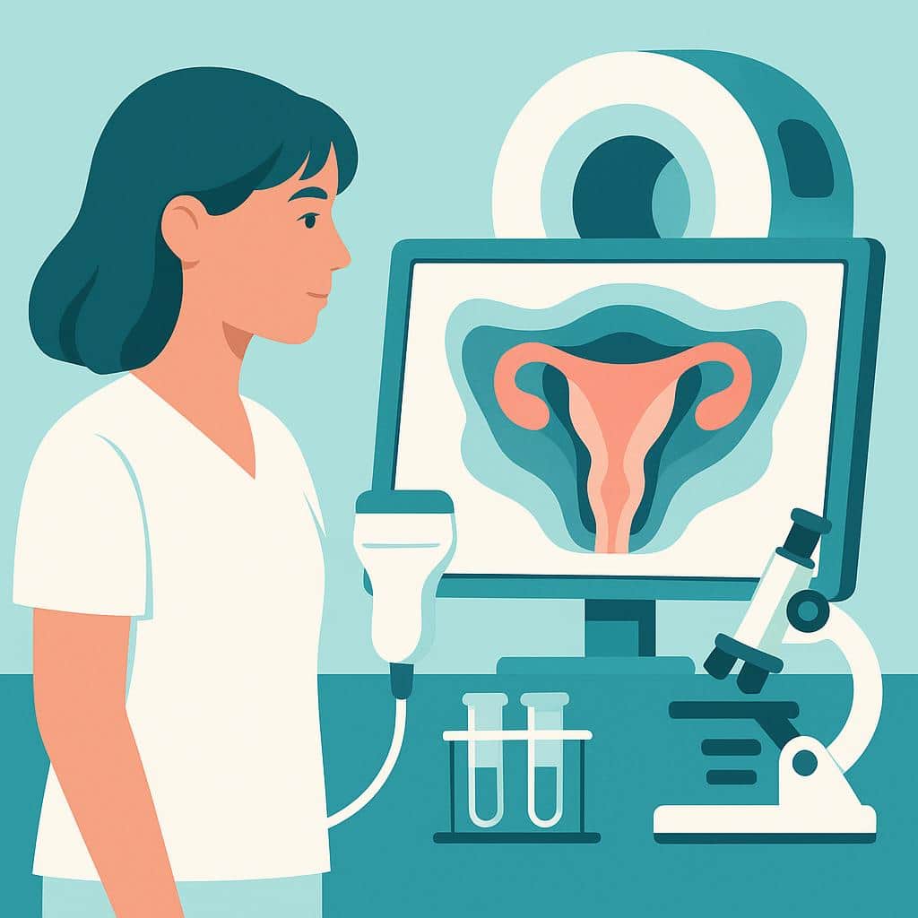
If you’ve been told “it might be adenomyosis,” your next question might be How do doctors actually diagnose adenomyosis—and how certain can we be without surgery?
The frustrating truth is that adenomyosis doesn’t have a single perfect, widely available, non-invasive “yes/no” test. Most real-world diagnoses are made by combining your symptoms, pelvic exam, and imaging—especially transvaginal ultrasound and sometimes MRI. Recent research also highlights why the condition is often missed: the accuracy of imaging can vary depending on the machine, the scan protocol, coexisting fibroids or endometriosis, and (very importantly) the experience of the person doing the scan.
This post synthesizes findings from multiple recent studies to help you understand what different tests can (and can’t) tell you—and how to use results to guide the next step in care.
The big picture: adenomyosis is usually an “evidence-based best fit” diagnosis
Historically, adenomyosis was only confirmed after a hysterectomy by looking at uterine tissue under a microscope. That’s still considered a “gold standard,” but it’s not a practical diagnostic pathway for most people who want symptom relief, fertility options, or uterine-sparing treatments. Modern care increasingly relies on imaging-based diagnosis, and the most patient-relevant shift is this: adenomyosis is now often treated as a condition you can presumptively diagnose well enough to guide management, even without tissue confirmation.
That said, imaging is definitely not infallible. A major review focusing on transvaginal sonography emphasized that reported sensitivity and specificity vary widely across studies—meaning your result can depend on where you’re scanned and how the ultrasound is performed. Another study in an IVF population drives this home: when women with recurrent implantation failure had a dedicated “expert” ultrasound, adenomyosis was reported far more often than on standard clinic scans, suggesting under-detection is real in routine settings.
First-line test: transvaginal ultrasound (TVS)
For most patients, transvaginal ultrasound (TVS) is the starting point because it’s accessible, non-invasive, relatively low-cost, and can show most characteristic patterns of adenomyosis. Across modern frameworks (including standardized reporting approaches), clinicians look for a constellation of findings rather than one single sign because there is a range of types of adenomyosis.
What your ultrasound report might mention (and what it means)
Different studies describe overlapping features that tend to show up in adenomyosis, including:
- Subendometrial echogenic lines/striations (bright lines or streaks near the lining) and myometrial heterogeneity (muscle looks “patchy” rather than uniform)
- Myometrial cysts (small fluid-filled spaces in the uterine muscle)
- Adenomyoma (a more localized “mass-like” area of adenomyosis)
- Junctional zone (JZ) changes—especially when measured with 3D ultrasound, where the inner transitional muscle layer may look thickened or irregular
- Descriptive signs like a globular uterus or “question mark”–type uterine contour in some scanning approaches
In a 2025 clinical study that focused on junctional zone assessment, people with adenomyosis had measurably thicker JZ measurements than imaging controls. That supports why many specialist ultrasound protocols pay close attention to the JZ—particularly in infertility evaluations where subtle uterine factors may matter.
Why “normal ultrasound” doesn’t always end the story
Ultrasound is operator-dependent. One multi-center-style clinical comparison in women with recurrent implantation failure found that an expert TVS exam detected pelvic pathology in the majority of patients who were labeled “normal” on prior clinic ultrasounds—and the disagreement was especially striking for adenomyosis (and also deep endometriosis). Fibroids, uterine position, and the choice of views (for example, whether 3D coronal views are obtained) can all influence what is seen.
If your symptoms strongly fit adenomyosis but your scan is “unremarkable,” it may be less about you and more about the limits of that particular scan.
When MRI is added—and what it’s best for
MRI is often used when:
- Ultrasound is inconclusive
- The case is complex (for example, coexisting fibroids that make ultrasound interpretation harder)
- Your care team needs more detail for treatment planning (including some uterine-sparing procedures)
A recent ultrasound-focused review noted that MRI may be preferable in complex scenarios and that combining MRI with TVS can improve overall diagnostic confidence—particularly for ruling out adenomyosis in certain settings. In practice, many clinicians use MRI as a “problem-solver” test rather than a universal first step. Also, if the diagnostic path is also looking for evidence of multi-organ endometriosis and deep invasive disease, an MRI may be the first choice.
MRI “extent” may relate to symptoms, but it’s not a pain meter
One 2025 study explored whether MRI-based severity patterns align with pain (dysmenorrhea) and whether a blood marker (CA125) adds useful information. The main patient takeaway isn’t that MRI can perfectly predict pain—it can’t. Rather the more extensive uterine involvement on MRI, the more it may be associated with different symptom patterns and biomarker levels in some populations.
This matters because patients often hear, “Your MRI looks mild—so why do you hurt so much?” Imaging findings and pain don’t always match neatly. Adenomyosis pain is influenced by inflammation, nerve signaling, uterine contractility, and overlapping conditions (including endometriosis), not just lesion size.
Blood tests: CA125 is sometimes helpful context, not a diagnostic answer
CA125 is a blood test usually used in ovarian cancer monitoring. But many patients are offered this test for pelvic pain or gynecologic workups. It can be elevated in several benign conditions (including endometriosis and adenomyosis), but it is not specific for endo or adeno or for cancer. So, it is not a very good tool for accuracy of diagnosis and may lead to unwarranted concerns about cancer.
In a 2025 cohort study, higher CA125 levels were associated with dysmenorrhea in patients whose MRI suggested more extensive adenomyosis, but the overall ability of CA125 to distinguish who had pain and who didn’t was modest (accuracy only slightly better than chance). In other words: CA125 may add context in selected cases, but it does not reliably diagnose adenomyosis or explain symptoms by itself.
If your CA125 is elevated, it’s reasonable to ask what else could be contributing—and whether imaging findings support adenomyosis, endometriosis, fibroids, or other gyn or non-gyn inflammatory causes, including cancer. Parenthetically, CA19-9 is also used in the same way occasionally.
Ready for Accurate Adenomyosis Diagnosis?
Our specialists are here to help you understand your condition and explore your treatment options.
Schedule Your ScanNewer ultrasound add-ons: 3D imaging, Doppler, and elastography
3D ultrasound and Doppler (blood-flow mapping)
Advanced ultrasound techniques—especially 3D TVS and color/power Doppler—are increasingly used in specialty settings. A key theme across recent evidence is that these tools may improve diagnostic precision in certain contexts, such as better visualization of the junctional zone or helping differentiate adenomyosis from fibroids in some cases.
This doesn’t mean every patient needs 3D or Doppler, but it does mean that how the scan is performed can change the yield—particularly when earlier imaging hasn’t matched your symptoms.
Cervical elastography: a promising “adjunct” signal
A 2026 study explored shear-wave elastography—a technique that measures tissue stiffness during transvaginal ultrasound. Interestingly, women with adenomyosis showed higher stiffness measurements near the internal cervical os than controls, and these measures had moderately strong ability to distinguish groups in that study.
The most important patient-friendly interpretation: this is not a replacement for standard ultrasound or MRI, and it hasn’t yet been proven to improve real-world diagnostic accuracy in the messy situations many patients face (like adenomyosis plus endometriosis). But it’s an example of where the field is heading: adding measurable “tissue properties” to imaging patterns to reduce uncertainty.
The “future test” patients hope for: urine or blood biomarkers (miRNA)
Because adenomyosis symptoms overlap so much with endometriosis and other pelvic pain conditions—and because imaging can be imperfect—researchers are actively looking for non-invasive biomarkers.
A 2025 pilot study used machine-learning models on microRNA (miRNA) patterns in urine and serum and reported that certain miRNA “signatures” could separate adenomyosis from endometriosis and from controls in that small cohort. The most exciting aspect for patients is the concept: a simple urine test could someday help triage who needs MRI, who needs specialist ultrasound, and who might have overlapping disease.
But the study also illustrates why this is not ready for clinics: the adenomyosis sample was extremely small, and “perfect” accuracy in tiny datasets often reflects overfitting rather than a true breakthrough. For now, this is best viewed as promising early research—not something you should expect your doctor to offer or rely on.
If you also have endometriosis (or suspect you do), diagnosis can get harder
Many patients have both adenomyosis and endometriosis, and separating symptoms can be difficult. Imaging can also be more challenging: deep endometriosis may require structured mapping and specialized skill to detect well on ultrasound, and adenomyosis features can be subtle.
One reason this matters is communication: if you’ve been told “it’s just adenomyosis” but you have bowel symptoms, pain with sex, or pain that doesn’t track with bleeding, it’s reasonable to ask whether a deep endometriosis evaluation was specifically performed (and by whom). The IVF imaging study showing low agreement for both adenomyosis and deep endometriosis between routine and expert scans underscores that overlap conditions can be missed without targeted protocols.
Practical takeaways: how to use your test results
Here are focused questions that often lead to better answers and next steps (bring the ones that fit your situation):
- “Was my ultrasound done with an adenomyosis-focused protocol (and were MUSA-type features assessed)?” Morphological Uterus Sonographic Assessment is shorted to MUSA and includes the imaging features discussed in this article.
- “Did the report comment on junctional zone features, myometrial cysts/striations, or adenomyoma?” (MUSA)
- “Could fibroids (or my uterine position) be limiting what ultrasound can see—would MRI add clarity?”
- “Do my symptoms suggest overlap with endometriosis, and was deep endometriosis specifically evaluated?”
- “If imaging is borderline, what would change management: trying medical therapy first, repeating imaging with an expert, or getting MRI?”
- “If my CA125 (or CA19-9) is elevated, what are the likely explanations in my case—and does it change anything we do?”
What we still don’t know
Even with better imaging standards and new technology, several gaps remain:
First, imaging performance varies across settings. Reviews of TVS show wide ranges in accuracy across studies, and real-world comparisons suggest expertise can dramatically change detection—especially for adenomyosis and deep endometriosis.
Second, symptom severity doesn’t map perfectly to what imaging shows. Studies linking MRI “extent” and biomarkers like CA125 or CA19-9 to pain find associations that are real but not strong enough to predict an individual person’s experience.
Third, many newer tools (like cervical elastography and miRNA testing) are promising but not yet validated in large, diverse populations—especially not in the common scenario of overlapping adenomyosis and endometriosis.
Finally, there isn’t a single universally agreed pathway for every patient. The best diagnostic plan is often tailored: your goals (pain relief, bleeding control, fertility), your risk factors, your prior imaging quality, and whether complex overlap disease is suspected should all influence what happens next.
Adenomyosis diagnosis is often less about finding one definitive test—and more about getting the right imaging, in the right hands, interpreted in the context of your symptoms. If your story and your results don’t line up, it’s a signal to ask for a more specialized or sophisticated look.
References
An, Zhang, Yun et al.. Perspectives in transvaginal sonography for the diagnosis of adenomyosis. Frontiers in Medicine. 2025. PMID: 40612584 PMCID: PMC12224653
Su, Huang, Wang et al.. Combined magnetic resonance imaging with serum CA125 for dysmenorrhea in adenomyosis. Scientific Reports. 2025. PMID: 41309741 PMCID: PMC12660971
Kupec, Wittenborn, Kuo et al.. Urine and Serum miRNA Signatures for the Non-Invasive Diagnosis of Adenomyosis: A Machine Learning-Based Pilot Study. Diagnostics. 2025. PMID: 41374393 PMCID: PMC12691541
Selntigia, Russo, Farsetti et al.. Expert transvaginal ultrasound is determinant for diagnosing pelvic conditions after recurrent implantation failure in IVF. European Journal of Obstetrics & Gynecology and Reproductive Biology: X. 2025. PMID: 41439198 PMCID: PMC12719194
Berbecaru, Zorilă, Istrate-Ofiţeru et al.. Adenomyosis-Modern Techniques for Ultrasound and Histo-Pathological Diagnosis of the Endo-Myometrial Junction Zone Changes. Journal of Clinical Medicine. 2025. PMID: 41464646 PMCID: PMC12733583
Kurt, Kurt, Duran Kaymak et al.. Evaluation of the Relationship Between Adenomyosis and Cervical Elastography Parameters. Journal of Clinical Medicine. 2026. PMID: 41753063 PMCID: PMC12942504
Quick Answers
Why do I have painful urination and pelvic cramping between periods?
Painful urination with pelvic cramping between periods can happen when the bladder, ureters, uterus, pelvic floor, or nerves are being irritated—sometimes in a way that still follows a subtle cycle pattern even if you’re not actively bleeding. Endometriosis can contribute by affecting the bladder wall or tissues around the bladder and ureters, and symptoms don’t have to include visible blood in the urine. Importantly, urinary tract endometriosis isn’t always “obviously urinary,” and ureter involvement can be quiet while still significant, which is why we take these symptoms seriously.
These symptoms can also come from conditions that overlap with (or mimic) endometriosis, such as bladder pain syndrome/interstitial cystitis, pelvic floor overactivity, adenomyosis-related uterine cramping, or other pelvic pain drivers. In our evaluation process, we focus on your full flare pattern—what triggers it, how it relates to your cycle, and whether urine tests have been repeatedly negative—then use targeted exam and the right imaging (often expertly interpreted ultrasound and/or MRI) to map what’s actually going on.
If you’re noticing recurring burning with urination, pressure, cramping, or symptoms that predictably flare mid-cycle or before your period, that pattern is useful diagnostic information—not something to dismiss. You can explore our urinary symptom and diagnostic evaluation resources to see how we approach “UTI-like” symptoms with negative cultures, and you’re welcome to reach out to schedule a consultation so our team can help you sort out whether this is bladder/ureter endometriosis, a look-alike condition, or a combination.
Which adenomyosis symptoms most affect daily life?
Adenomyosis symptoms that most disrupt quality of life usually come from two main issues: how the uterus bleeds and how it hurts. Many patients describe heavy or prolonged periods that interfere with work, school, travel, and sleep—sometimes with flooding, frequent pad/tampon changes, and fatigue that can follow significant blood loss. Severe period pain (often more than “normal cramps”) is also common, and it can feel deep, aching, or pressure-like, sometimes accompanied by an enlarged, tender uterus and a sense of pelvic heaviness or bloating.
Outside of the period itself, adenomyosis can contribute to chronic pelvic pain, pain with sex for some people, and bowel or bladder discomfort—especially when symptoms flare around the menstrual cycle. It can also overlap with endometriosis, and when both are present symptoms may intensify or become harder to tease apart. If your day-to-day life is being shaped by bleeding, pain, pressure, or fertility stress, our team can help you sort out whether adenomyosis, endometriosis, fibroids, or more than one condition may be driving the pattern—and what next-step options make sense for your goals.
Why do I look pregnant from bloating with constant pelvic pressure?
Feeling so bloated you “look pregnant” along with constant pelvic pressure usually points to more than simple gas—often it’s a pelvic condition creating inflammation, swelling, or a sense of bulk. Endometriosis can irritate the bowel and pelvic lining, trigger scarring that tethers organs, and create the classic “endo belly” sensation that comes and goes (sometimes not perfectly cyclical). Pelvic pressure can also happen when endometriosis involves deeper tissues or nearby organs like the bladder, ureters, or rectum.
Just as important: these symptoms can be driven by endometriosis neighbors or coexisting conditions, especially adenomyosis and fibroids, which can make the uterus feel heavy, full, or “bulky” and add pressure on the bladder and bowel. Ovarian cysts and other benign pelvic findings can contribute, and IBS-like bowel sensitivity can overlap so closely that symptoms alone don’t reliably sort out what’s causing what. Our team focuses on mapping the full picture—uterus, ovaries, bowel, bladder, and pelvic support structures—so treatment targets the true driver(s), not just the most obvious diagnosis.
If this pressure/bloating is persistent, worsening, or changing your ability to eat, move your bowels, or urinate comfortably, it’s a strong reason to pursue a deeper evaluation rather than being told it’s “normal.” You can explore our educational content on bowel symptoms, bladder symptoms, and overlapping conditions, and reach out to schedule a consultation so we can review your history, imaging, and symptom pattern and outline a plan aimed at lasting relief.
Why does ovarian cyst pain keep coming back?
Ovarian cyst pain can feel “recurrent” for a few different reasons. Some cysts are functional (they form with ovulation and then resolve), so the pain returns in a similar spot month after month even though it’s not the exact same cyst. In other cases, the cyst itself can come back or persist—especially if it’s an endometrioma (an ovarian cyst caused by endometriosis), which can behave differently than a simple cyst and may be associated with deeper pelvic disease.
Another common reason is that the cyst isn’t the whole story: endometriosis on the pelvic sidewall, uterosacral ligaments, bowel, bladder, or around the ovary can irritate the same nerves and tissues and make it feel like “my cyst is back” when the driver is actually inflammatory disease nearby. Adhesions (scar-like bands) can also tether the ovary and cause recurring pulling or sharp pain, even when imaging doesn’t show a large cyst. If your pain cycles, escalates, returns quickly after a “normal” ultrasound, or keeps recurring despite prior treatment, our team can help you map the pattern, interpret imaging with an endometriosis lens, and decide whether targeted evaluation and—when appropriate—excision surgery is the next best step.
Why do I get low back and leg pain during my period?
Low back and leg pain that predictably flares with your period can happen when pelvic inflammation irritates pain pathways that “refer” into the back, hips, buttocks, and down the leg. In some patients, endometriosis can be part of that story—either indirectly (pelvic inflammation and scarring increasing pressure and sensitivity around nearby nerves) or more directly if disease is affecting areas close to major nerves.
When period-related leg pain resembles sciatica—deep buttock pain, tingling, burning, or pain radiating down the back of the thigh—it raises the possibility of endometriosis-related sciatic irritation or pelvic floor involvement (often described as piriformis-type pressure on the nerve). These symptoms may start before bleeding, peak during the period, and linger afterward, and in more significant cases can be associated with weakness or changes in walking.
Because back and leg pain can also come from the spine, hips, or muscles, the key is the pattern and the full symptom “constellation,” including pelvic pain, bowel/bladder symptoms, or pain with sex. Our team can help you sort out whether your pain fits an endometriosis/adenomyosis pattern and, if needed, plan next-step evaluation such as targeted imaging and a strategy focused on lasting relief rather than temporary suppression.